On February 10, 2026, the National Institutes of Health (NIH) announced what scientists are calling a “paradigm shift” in vision science: the first ever subcellular resolution digital twin of a human eye cell.
Unlike a static 3D model, this digital twin is a data driven, AI powered biological replica that allows researchers to observe how a living cell develops, ages, reorganizes, and ultimately fails in real time.
The breakthrough could dramatically accelerate research into blindness, drug development, and personalized medicine.
The POLARIS Breakthrough
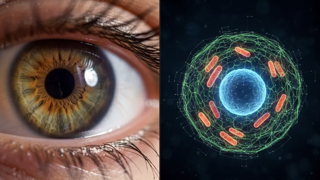
The project focuses on Retinal Pigment Epithelial (RPE) cells, the support cells that sit behind the retina. Often described as the “janitors” of the eye, these cells:
- Recycle waste from light-sensing photoreceptors
- Deliver nutrients from the bloodstream
- Maintain strict internal organization known as “polarity”
When RPE cells lose this top to bottom organization and die, it can lead to Age Related Macular Degeneration (AMD)
the leading cause of blindness in people over 50.
To tackle this, researchers at the National Eye Institute, led by Kapil Bharti, developed an AI system called POLARIS (Polarity Organization with Learning based Analysis for RPE Image Segmentation).
How the Digital Twin Was Built
1. Reprogramming Human Cells
Because living retinal cells cannot easily be extracted from patients, scientists used induced pluripotent stem cells (iPSCs). These are ordinary skin or blood cells reprogrammed into RPE cells, the same cells that fail in macular degeneration.
2. Industrial-Scale 3D Imaging
The team analyzed 1.3 million RPE cells using automated confocal microscopy:
- Nearly 4,000 hig -resolution 3D imaging fields
- Fluorescent tagging of nuclei, mitochondria, and cytoskeletal structures
- Voxel level precision (3D pixels instead of flat 2D images)
This produced one of the most detailed cellular datasets ever assembled.
3. The POLARIS AI Engine
POLARIS assigns a label to every voxel inside a cell, identifying the precise 3D location, volume, and boundaries of each organelle.
For a general reader, think of it this way:
POLARIS doesn’t just see the cell’s “face.” It sees the plumbing, the wiring, and the structural beams the cytoskeleton in full 3D.
It can detect when the cell’s internal “foundation” begins to shift long before the entire structure collapses.
Instead of a snapshot, researchers now have a living architectural blueprint.
4. Mapping Cellular Polarity
Healthy RPE cells are polarized:
- Apical side: Faces light sensitive cells and removes waste
- Basal side: Faces the blood supply to absorb nutrients
In disease states, this internal orientation breaks down.
The digital twin continuously tracks the spatial coordinates of mitochondria, nuclei, and structural proteins. When these components begin drifting from their predictable layout, POLARIS flags it even before visible degeneration occurs.
This shift is known as a cell state transition the moment a cell moves from a healthy, polarized configuration into a diseased, disorganized one.
Why This Matters for Blindness Treatment
Early Warning System
Traditional microscopy shows what a cell looks like now.
The digital twin models how it is changing over time. Scientists can pinpoint the exact stage when a cell begins transitioning toward failure potentially months before permanent damage.
Virtual Drug Testing Targeting Cell State Transitions
The most powerful application may be virtual drug testing.
Instead of simply observing whether a drug keeps a cell alive, researchers can now measure whether a therapy prevents a cell state transition stopping the shift from healthy polarization to diseased disorganization.
In practical terms, the AI can simulate:
- Does Drug X maintain proper organelle positioning?
- Does it push drifting mitochondria back into healthy alignment?
- Does it halt the transition into a non polarized state?
By modeling these changes digitally, researchers can prioritize the most promising therapies before moving into lengthy animal or human trials.
Toward Personalized Digital Twins
Because the model is built from patient derived stem cells,
it opens the possibility of individualized eye cell twins.
In the future, physicians could:
- Generate a digital twin from a patient’s own cells
- Predict the rate of degeneration
- Simulate multiple treatments
- Choose the therapy most likely to preserve vision
Beyond the Eye: The Rise of Organ Digital Twins
The eye breakthrough represents the first subcellular resolution success, but digital twin research is expanding rapidly across medicine.
The Heart
Researchers at Johns Hopkins University and King’s College London have created thousands of anatomically precise virtual hearts.
Surgeons use these twins to rehearse cardiac ablations for arrhythmias before operating on real patients, improving accuracy and outcomes.
The Liver
NIH funded multiscale liver twins are being used to predict drug-induced liver injury, including acetaminophen toxicity potentially reducing the need for animal testing.
The Brain
Digital brain models are helping neurologists simulate seizure origins in epilepsy patients, allowing for more precise surgical planning.
The Placenta
Emerging placental twins use ultrasound and MRI data to predict complications such as pre eclampsia and complex labor.
The Next Frontier: Whole Body Medical Avatars
In 2026, NIH workshops have begun focusing on modeling inter organ communication, how a digital heart interacts with digital lungs, kidneys, and brain.
The long term goal is a full body digital twin: a lifelong medical avatar capable of predicting disease risk, simulating treatment response, and tracking aging at the cellular level.
A Blueprint for the Future of Medicine
According to Dr. Bharti, the RPE digital twin serves as a blueprint for studying diseases where cellular organization breaks down including cancer and neurodegenerative disorders like Alzheimer’s.
The shift marks a broader transformation in medicine: moving from observing damage after it occurs to predicting and potentially preventing it before symptoms appear.
For vision research and beyond, the digital twin era has officially begun.